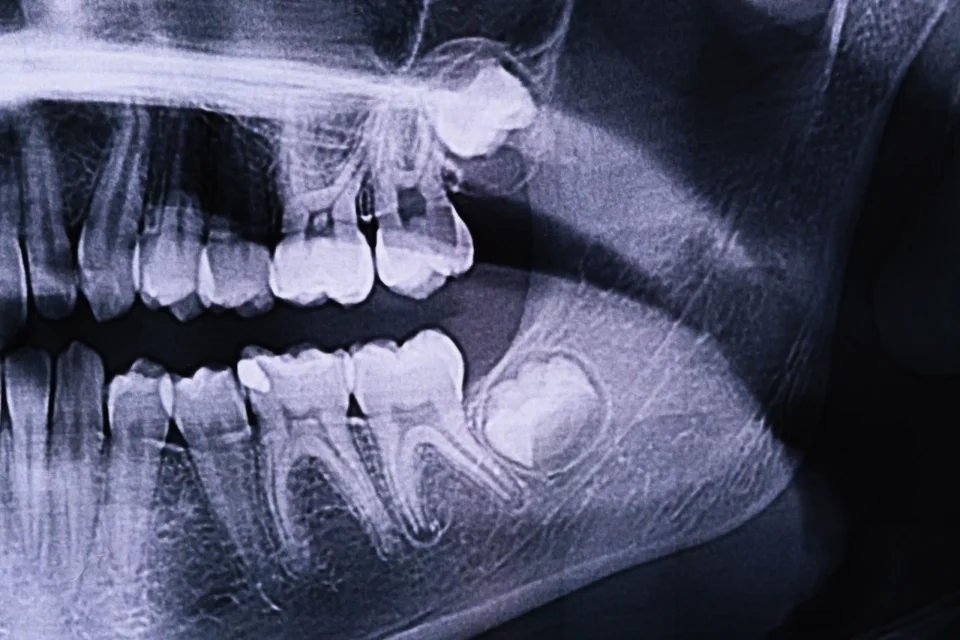
Root canal treatment is a dental procedure often necessary when a tooth’s pulp, which contains nerves and blood vessels, becomes infected or inflamed. This may happen due to a tooth crack or chip, extensive decay, faulty crowns, or repeated dental treatments. Dentists utilize X-rays as a critical diagnostic tool in such cases to evaluate the severity of the damage and devise treatment strategies. Furthermore, let’s delve into the details of understanding an X-ray of a tooth that needs a root canal.
Identifying the Signs on an X-Ray of a Tooth that Needs Root Canal
When looking at an X-ray of a tooth that requires root canal treatment, dentists look for many vital signs. These include:
- Dark Spots at the Tip of the Tooth’s Roots: These spots signify an infection in the bone near the tooth’s end, indicating that the pulp is diseased or dead.
- Bone Loss Around the Root: This is a sign of a long-term infection that may necessitate a root canal.
- Changes in the Root Canals: An X-ray may show that the root canals are enlarged or shaped irregularly, a sign of an infection.
- Visible Signs of Decay: Deep decay extending to the pulp chamber clearly indicates that root canal therapy may be necessary.
The Process of Root Canal Treatment
Step 1: Diagnosis with an X-Ray
The first step in the root canal process is a thorough examination, including an X-ray of the tooth that needs a root canal. Moreover, this X-ray allows the dentist to assess the shape of the root canals and determine whether there are any signs of infection in the surrounding bone.
Step 2: Anesthesia and Access
Once a root canal is deemed necessary, the dentist numbs the tooth’s surrounding area with local anesthesia. Then, they will drill an access hole into the tooth to allow access to the pulp chamber and root canals.
Step 3: Cleaning and Shaping
The dentist removes the infected pulp tissue and utilizes small instruments to clean and shape the root canals. This procedure is critical for eliminating all bacteria and debris and avoiding reinfection.
Step 4: Filling and Sealing
After the canals are cleaned and dried, they are filled with a biocompatible material, usually a rubber-like material called gutta-percha. Then, the dentist seals the access hole with a temporary or permanent filling.
Step 5: Restoration
Finally, the tooth will need a crown or other restoration to protect and restore it to full function.

Post-Root Canal Care
After a root canal treatment, it’s normal to experience some discomfort or mild pain. You can usually manage this with over-the-counter pain relievers. In order to ensure the longevity of the treated tooth, it’s critical to maintain proper oral hygiene practices, which include brushing, flossing, and routine dental checkups.
Get to the Root of the Matter: Your FAQs on X-Rays of Teeth Need Root Canals Answered!
What does a tooth look like when it needs a root canal?
A tooth that needs a root canal may not always show visible signs, but when it does, these can include:
- Discoloration: The tooth may become darker or discoloured due to internal damage.
- Swelling and Tenderness in the Gums: The area around the affected tooth may be swollen or tender.
- Visible Decay or Damage: There may be obvious signs of decay or a crack in the tooth.
- A Pimple on the Gums: Sometimes, a small pimple-like bump, known as a dental abscess, can form on the gums near the affected tooth.
Does a failed root canal show on an X-ray?
Yes, dentists can often detect a failed root canal on an X-ray. Signs may include:
- Persistent Dark Spots or Lesions at the Root Tips: This indicates ongoing infection or an abscess.
- Inadequate Filling Material: The dentist may need to fill the root canals more adequately, or there may be voids in the filling material.
- Recurrent Decay: New decay around the treated tooth can be visible.
- Further Bone Loss: Continued bone loss around the root tip suggests an ongoing problem.
Can tooth root infection be seen on an X-ray?
Yes, tooth root infection is often visible on an X-ray. The infection can manifest as dark spots or lesions at the root tips, indicating the presence of an abscess or bone loss due to the infection. Also, changes in the appearance of the root canal itself, such as enlargement or irregular shape, can be a sign of infection. However, in the early stages, an infection might not always be visible on an X-ray, so clinical symptoms and examination are crucial for diagnosis.
Final Thoughts
Understanding the X-ray of a tooth that needs a root canal is crucial in diagnosing and treating dental problems effectively. Sunshine Dentistry in Richmond Hill, Ontario, employs cutting-edge technology and techniques to provide our patients with the best care possible. If you are experiencing dental pain or discomfort, please do not hesitate to contact us. Early intervention can often save a tooth and prevent more complex treatments. Call us today!


