Root canals are a standard dental procedure to save a severely damaged or infected tooth. A root canal X-ray is essential for diagnosing and performing this procedure. This diagnostic method provides detailed images of the tooth’s interior, allowing dentists to assess the extent of damage, plan treatment, and track the healing process. Furthermore, in this blog, we will discuss the significance of X-rays and their role in the treatment process and answer frequently asked questions about them.
What is a Root Canal X-Ray?
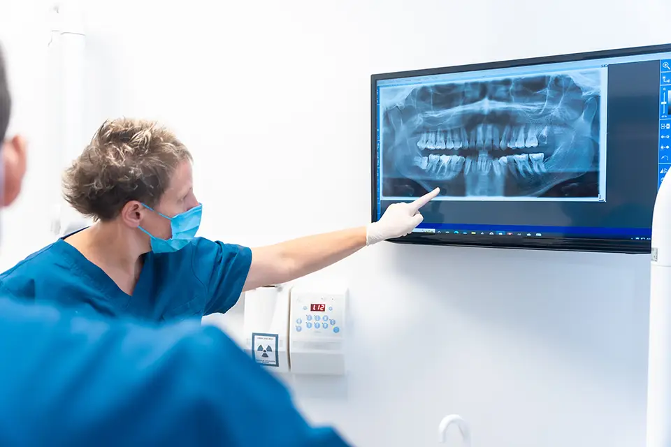
A root canal X-ray is a specialized dental imaging technique that takes detailed images of a tooth’s internal structure, including the root canals and surrounding bone. These X-rays assist dentists in detecting issues such as infections, fractures, or previous treatments that would not be visible during a routine dental exam. They are essential for planning and executing a successful treatment because they provide a clear view of the tooth’s root and surrounding tissues.
Why are Root Canal X-rays Important?
Root canal X-rays are necessary for a variety of reasons.
- Accurate Diagnosis: They assist dentists in determining the precise location and extent of tooth damage or infection, which is critical when determining the need for a root canal procedure.
- Treatment Planning: X-rays provide a detailed view of the tooth’s anatomy, allowing the dentist to accurately plan the procedure and ensure that all infected areas are treated effectively.
- Monitoring Progress: Dentists use X-rays during and after treatment to ensure that the infection is healing and that there are no complications.
- Preventing Future Issues: They can detect potential problems, such as additional canals or fractures, that would complicate treatment if left unnoticed.
The Root Canal X-Ray Process
The root canal X-ray procedure is straightforward and painless. Here’s what you can expect:
- Initial Examination: Before beginning the procedure, the dentist will perform an initial x-ray to assess the tooth’s condition. This aids in diagnosing the issue and planning the treatment.
- During the Procedure: X-rays may be taken during the root canal to ensure the cleaning and filling proceed correctly. This step is critical to ensure the removal of all infected tissue.
- Post-Treatment Monitoring: After the procedure, the dentist takes additional X-rays to monitor the healing process and ensure that the tooth and surrounding bone recover properly.
Types of Root Canal X-Rays
There are various types of x-rays used in root canal treatments, each with a specific purpose:
- Periapical X-rays: These provide a complete view of the tooth, from the crown to the root tip, including the surrounding bone. Dentists frequently use them to detect infections and evaluate the health of the root and bone.
- Bitewing X-rays: These show both the upper and lower teeth in one area of the mouth and are commonly used to detect tooth decay or changes in bone density caused by gum disease.
- Panoramic X-Rays: These offer a comprehensive view of the mouth, including all teeth, jaws, and sinuses. They help detect overall dental health problems and develop extensive treatment plans.

How to Prepare for a Root Canal X-Ray
Preparing for an X-ray is simple. There is no need for any special preparation, but here are some tips to ensure the process runs smoothly:
- Inform Your Dentist: Inform your dentist if you have any dental appliances, such as braces or dentures, that could interfere with the X-ray.
- Remove Jewelry: You should remove metal objects, such as earrings or necklaces because they may obstruct the X-ray image.
- Stay Still: It is critical to stay still during the X-ray to avoid blurring the image.
Benefits
X-rays provide numerous advantages that contribute to the success of the treatment:
- Precision in Diagnosis and Treatment: They enable precise identification of the problem area, resulting in more effective treatment.
- Early Detection of Complications: X-rays can detect issues such as additional canals or fractures early on. Moreover, they reduce complications during and after the procedure.
- Efficient Monitoring: They aid in tracking the healing process, ensuring that the tooth and surrounding bone recover as expected.
FAQs
1. Are X-rays safe?
Yes, they are safe. The radiation exposure is minimal, and dentists take precautions to protect patients.
2. How often will I need a root canal X-ray?
To ensure successful results, you typically need one before, during, and after the root canal treatment.
3. Do X-rays hurt?
No, taking an X-ray is a painless procedure. You might feel slight discomfort from holding the film or sensor in your mouth.
A root canal X-ray is valuable for accurately diagnosing and treating dental issues. It provides critical information for planning and executing a successful root canal procedure. Furthermore, if you suspect you need a root canal or have been advised to get one, rest assured that root canal X-rays will ensure the best possible outcome for your dental health. For more information or to schedule a consultation, contact Sunshine Dentistry in Richmond Hill, Ontario, today!